1/4ページ
ダウンロード(539.9Kb)
バイオ医薬品の多様性と純度分析の世界標準ツール
新方式キャピラリー電気泳動 『Maurice(モーリス)』
バイオ医薬品の多様性(バリアント)分析および
純度分析の最新プラットホーム。
等電点電気泳動(cIEF)とSDS電気泳動(CE-SDS)
を一台で簡単に行います。
【特長】
New!:等電点電気泳動で分離後のサンプル分取 (MauriceFlex)
New!:5.5分(還元処理IgG) ~ 8分(非還元処理IgG) での迅速な泳動検出 (Maurice Turbo CE-SDS)
●新方式の一体型キャピラリーカートリッジ
●cIEFとCE-SDSの切り替えはカートリッジの交換のみ
●キャピラリーのセットアップと洗浄工程を完全自動化
●オートサンプラー内蔵
●オンボードミキシング内蔵(オプション)
●cIEFモードでは高感度ネイティブ蛍光検出利用可能
●21 CFR Part 11に準拠
関連メディア
このカタログについて
| ドキュメント名 | 新方式キャピラリー電気泳動 Maurice |
|---|---|
| ドキュメント種別 | ホワイトペーパー |
| ファイルサイズ | 539.9Kb |
| 登録カテゴリ | |
| 取り扱い企業 | プロテインシンプル ジャパン株式会社 (この企業の取り扱いカタログ一覧) |
この企業の関連カタログ

このカタログの内容
Page1
Spotlight
A Novel Platform for icIEF Fractionation
of Antibody Charge Variants
While imaged capillary isoelectric focusing Experimental Methods
electrophoresis (imaged cIEF or icIEF) has become the
The stepwise fractionation workflow on MauriceFlexTM
method of choice for monitoring charge variant levels is
of biotherapeutics, the in-depth characterization of illustrated below:
charge variants has been carried out mostly by mass
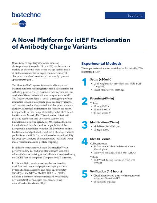
spectrometry (MS). 1 Setup (~30min)
• Load reagents (kit provided) and NIST mAb
The MauriceFlexTM system is a new and innovative (1 mg/mL)
Maurice platform featuring icIEF-based fractionation for • Insert MauriceFlex cartridge
collecting protein charge variants, enabling downstream
analysis of these variants with techniques such as MS.
The fractionation utilizes a special cartridge to perform 2 Focusing (45min)
isoelectric focusing to separate protein charge variants, Voltage
and once focused and separated, the charge variants are • 10 min @500 V
eluted via chemical mobilization for fraction collection. • 10 min @1000 V
Compared to ion-exchange chromatography (IEX)-based • 25 min @1500 V
fractionation, MauriceFlexTM fractionation is fast, with
pI-based resolution, and overcomes some of the
limitations of direct coupled cIEF-MS, such as the need 3 Mobilization (25min)
for a dedicated interface and incompatibility of the • Mobilizer: 5 mM NH4Ac
background electrolytes with the MS. Moreover, offline • Voltage: 1000V
fractionation and potential enrichment of charge variants
pooled from multiple fractionations offer more flexibility
for mass spectrometry characterization, including intact 4 Elution (20min)
mass, reduced mass and peptide mapping.
Collect fraction
In addition to fraction collection, MauriceFlexTM can • 36 fractions at 25 second/fraction on a
96-well plate
perform routine CE-SDS and cIEF analysis using the
• Each well contains 30 uL 5 mM NH4Ac
relevant Maurice cartridges, and all data is analyzed using
the 21CFR Part 11 compliant Compass for iCE software. Voltage
• 1000 V (off during transition from well
In this spotlight, we demonstrate the fractionation to well)
workflow and intact and peptide mapping analysis
by liquid chromatography and mass spectrometry
5 Verification (4-5 hours)
(LC-MS) on the NIST mAb (RM 8761 from NIST),
which is a common reference standard for assessing • Check identify and purity of fractions with
analytical Maurice icIEF
new analytical technologies for characterizing
monoclonal antibodies (mAbs). • 16 fractions checked
Page2
For intact mass, the fractions were analyzed directly. A
0.25 mg/mL
For peptide mapping, the charge variant fractions from
A2 A1 M B1 B2
10 fractionation runs were pooled, lyophilized on a 30,000
SpeedVac, and reconstituted prior to tryptic digestion.
Variant Relative
The digested samples were lyophilized and reconstituted 25,000 Abundance
in 40 µL 5 mM ammonium acetate solution. B2 0.95
20,000
B1 8.5%
The LC-MS characterization was performed with a 15,000
M 58.4%
Thermo ScientificTM Vanquish UHPLC coupled to a Q
10,000 A1 25.7%
ExactiveTM HF Hybrid Quadrupole-Orbitrap™ mass
A2 6.5%
spectrometer. Reverse phase LC separation was used 5,000
for intact mass analysis and peptide mapping with 0
appropriate gradients of 0.1% formic acid in water and 8.7 8.8 8.9 9 9.1 9.2 9.3 9.4
acetonitrile at 0.3 mL/min. The injection volume was pI
10 µL for both intact and peptide mapping analysis. The
data were analyzed using BioPharma Finder 4.1 software. B
1 mg/mL
20,000
A2 A1 M B1 B2
Results
The method for fractionation of the NIST mAb sample 15,000
using the MauriceFlex cIEF fractionation cartridge can
be developed quickly based on the analytical method
10,000
on a Maurice cIEF cartridge. FIGURE 1 shows profiles
of analytical icIEF and fractionation focusing runs of
NIST mAb. Five charge variants (B2, B1, M, A1, A2) were 5,000
identified for NIST mAb and their relative abundances
were obtained (FIGURE 1A). Note that fractionation 0
separation with cIEF fractionation cartridge is designed 8.6 8.8 9 9.2 9.4
for maximizing the yield of the fraction collection, and for pI
this purpose, a higher concentration (1 mg/mL) of NIST FIGURE 1. icIEF separation of NIST mAB charge variants with the Maurice
cIEF cartridge (A) and MauriceFlexTM cIEF Fractionation cartridge (B). Both
mAb was loaded. While this resulted in apparently lower electropherograms show the same number of charge variants. The relative
resolution with overlapped peaks (FIGURE 1B) when abundance (%) of each variant was calculated from the peak areas in the
compared to charge separation with the regular Maurice analytical run (A) and listed in the table insert.
cIEF cartridge (FIGURE 1A), the same number of charge
variants were detected, as seen in FIGURE 1B, and were
well separated.
As shown in the workflow, the mobilization and elution 40,000
steps take 45 minutes in total when collecting 36 fractions 20,000 Reference Standard
0
on a 96-well plate. The Compass for iCE software has 40,000
a peak prediction feature that provides an estimated 20,000 #10 (65% B2, 35% B1)
0
range of wells that contain the eluted charged variants. 40,000
20,000 #11 (100% B1)
Alternatively, a fluorescence plate reader can be used to 0
40,000
select the wells that contain the most abundant charge 20,000 #14 (100% M)
variant. For NIST mAb, a total of 16 wells of fractions were 0
40,000
selected, and their identity and purity were verified with 20,000 #17 (100% A1)
0
analytical Maurice icIEF. Among the 16 fractions analyzed, 40,000
20,000 #21 (100% A2)
12 were found to contain the charge variants. 0
8.4 8.6 8.8 9 9.2 9.4 9.6
FIGURE 2 shows icIEF electropherograms of fraction wells pI
containing individual charge variants with the highest FIGURE 2. Verification of charge variants of representative fractions for
purity. As shown, except for low abundant B2 (0.9%) at 65% identity and purity by Maurice icIEF analytical runs. The numbers assigned
represent the fraction numbers.
purity in fraction #10, fractions containing 100% purity for
the four other charge variants were obtained.
Fluorescence Fluorescence Fluorescence
Page3
1_25s_NIST_B2 NL: 3.91E5 1_25s_NIST_B2 #6192-6525 RT:5.219-5.500 AV:334 NL:
6.31E4 1_25s_NIST_B2 NL:
2.90E6
5.332 100 3093.8529
100 148455.08
100
3228.3434 148615.67
148293.41
B1
50 50 3374.9532 50 148046.48
148776.81
5.513 3621.8044 148154.77
147832.36 148936.20
2320.1353 4013.4516 4364.7178
0
2_25s_NIST_B1 NL: 8.33E5 0
2_25s_NIST_B1 #6192-6538 RT:5.219-5.500 AV:347 NL: 0
1.49E5 2_25s_NIST_B1 NL:
6.60E6
5.327 3091.1942 148323.80
100 100 100
148162.06
B2 3297.0919 148488.27
50 5.406 50 50 147957.50
3450.4218
5.458 3804.3165 148119.58 148647.80
2179.1503 4125.6578 148812.16
0 NL:
3_25s_NIST_M NL: 1.27E8 0 0
3_25s_NIST_M #6196-6631 RT:5.219-5.500 AV:436 3_25s_NIST_M NL:
1.30E7 5.93E8
5.247 3088.5136 148195.83
100 100 100
148360.19
M 3294.3404 148034.06
147832.55
50 50 3447.4749 50 148521.97
3800.9019 147983.95
148680.13
5.366 2352.1516 4239.9135
0 NL: 2.24E7 0
4_25s_NIST_A1 #6192-6606 RT:5.219-5.500 AV:415 NL: 0 NL:
4_25s_NIST_A1 1.85E6 4_25s_NIST_A1 7.28E7
5.262 100 3091.9231
100 100 148359.86
148520.52
148196.08
3297.8927
50 A1 147985.31 148680.31
50 3451.2775 50
5.309 148037.08 148835.75
3809.3392 148995.39
5.461 2318.6478 4122.2188 149157.33
0 0
5_25s_NIST_A2 #6192-6525 RT:5.219-5.500 AV:334 NL: NL:
5_25s_NIST_A2 NL: 3.43E5 0
5_25s_NIST_A2
4.83E4 2.32E6
3157.5700
100 5.343 148361.48
5.311 100 100
5.372 148199.25
3297.9091 148521.36
A2 147987.39
148683.70
50 50 3455.0008 50 148036.25
148842.23
3805.0788
2355.1556 4239.4999 148994.20
0 0 0
3.0 3.5 4.0 4.5 5.0 5.5 6.0 6.5 7.0 7.5 8.0 2000 2500 3000 3500 4000 4500 147000 147500 148000 148500 149000 149500 150000
RT (min) m/z Mass
FIGURE 3. LC-MS intact mass analysis of charge variants showing total ion
current (TIC) chromatograms (left), mass spectra of the peaks (middle) and
the deconvoluted spectra (right).
Peak Deconvoluted
Mass (Da) Mass Shift (Da) Modification
The LC-MS intact mass analysis on the high purity B2 148455.08 +259.25 2xC-term K
charge variant fractions are shown in FIGURE 3 and
B1 148323.80 +127.97 C-term K
the modifications identified from the deconvoluted mass
M 148195.83 0.00 G0F/G1F
spectra for each charge variants are summarized in
TABLE 1. The results are consistent with the established A1 148359.86 -164.03 Glycation
knowledge of PTMs of the charge variants of NIST mAb. A2 148361.48 -165.65 Glycation
TABLE 1. Summary of the identified modifications from intact mass analysis.
The mass shifts of each variants are relative to the G0F/G1F glycoform of
the M peak.
NL: 1.32E8 Peptide mapping is an indispensable tool for
22.98
100
90 characterizing the primary structure of biotherapeutics,
80 B1
70 and the capability of pooling charge variants from
60
50 11.56
1.22 multiple fractionation runs on MauriceFlexTM makes it
23.13
40
11.03 15.00
30 9.41 14.74
6.57 22.74 possible to enrich the charge variants for peptide mapping
7.52
20 23.22
4.94 17.80
10 17.38
1.97 4. 19.93 21.30 24.07 analysis. FIGURE 4 demonstrates the peptide mapping
1.05 53 25.12 28.79
0
2 4 6 8 10 12 14 16 18 20 22 24 26 28 30 results from samples pooled from 10 fractionation
RT (min) NL: 2.32E8
11.55 runs. As shown, the sequence coverage is above 90%
100
90
A1 22.97 for all variants except for the low abundant B (relative
80 9.41 11.04
70 7.52 14.98
14.72
19.92 abundance 0.9%) and heavy chain (HC) of the A2 peak.
60 6.56 21.27
9.69 17.82
50
40 18.46
8.96 22.18 23.12
30
1.22
20 17.38 23.21
10 4.93 Sequence Coverage %
8.
0 1.97 4.54 23
.68 16.80 24.38 25.17 27.96 29.74
0
2 4 6 8 10 12 14 16 18 20 22 24 26 28 Fraction LC HC
RT (min)
B2 84.0 58.9
FIGURE 4. Representative chromatograms of the peptide mapping of B1 and
A1, and sequence coverage of Light Chain (LC) and Heavy Chain (HC) of all B1 94.4 92.4
charge variants. The charge variant samples were pooled from 10 M 95.3 94.0
fractionation runs.
A1 95.3 92.2
A2 93.9 73.8
Relative Intensity Relative Intensity Relative Intensity
Relative Intensity
Relative Intensity
Page4
Conclusion
By using the NIST mAb sample with the icIEF • A single fractionation run provides sufficient charge
fractionation feature on the new MauriceFlexTM system, variant fractions for intact mass analysis
we have demonstrated that: • With enrichment from pooling fractions from multiple
runs, charge variants can be analyzed with LC-MS
• High purity fractionation of charge variants can be peptide mapping
obtained in a single day
Bio-Techne® | R&D Systems™ Novus Biologicals™ Tocris Bioscience™ ProteinSimple™ ACD™ ExosomeDx™ Asuragen®
For research use or manufacturing purposes only. Trademarks and registered trademarks are the property of their respective owners.
STRY0297039_BBU_SP_MauriceFlex-Spotlight_KG


